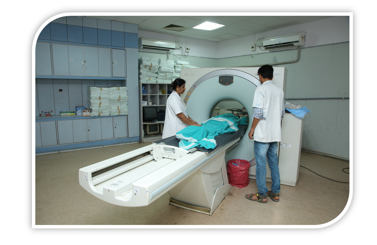
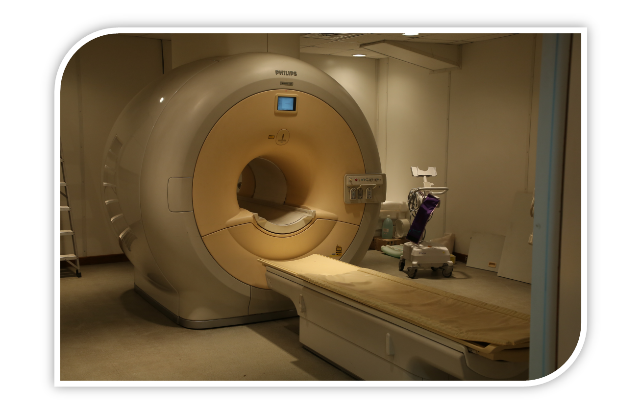
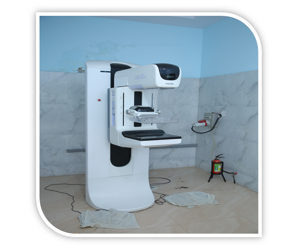
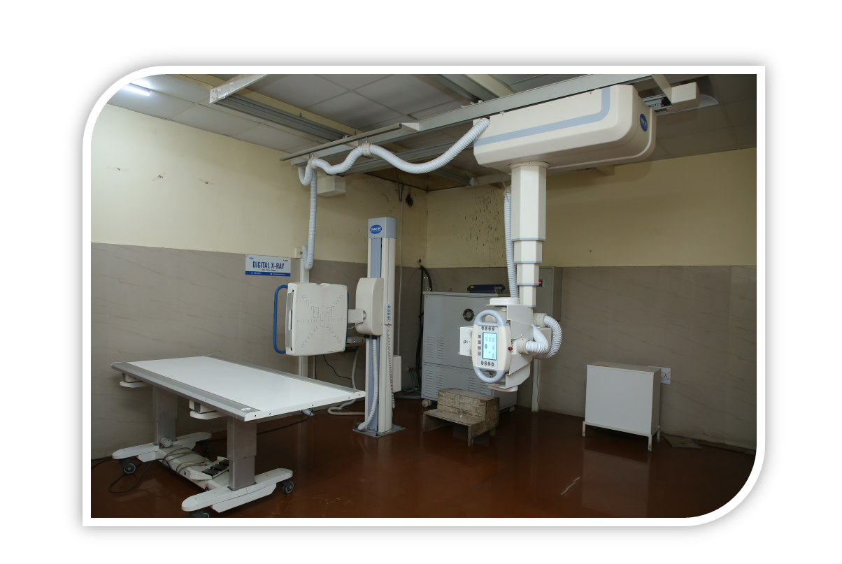
The Department of Radiodiagnosis from its inception, continues to play an important and significant role in the over all health care and academic activities of the Institute. The department is well equipped with various imaging equipments such as Digital X-Ray and fluroscopy machines, Ultrasound / colour Doppler machines, Mammography Unit, Whole body CT Scanners as well as 1.5 Tesla MRI equipment, Digital Subtraction Angiography. PACS is present for efficient reporting.
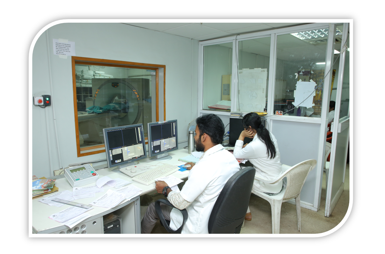
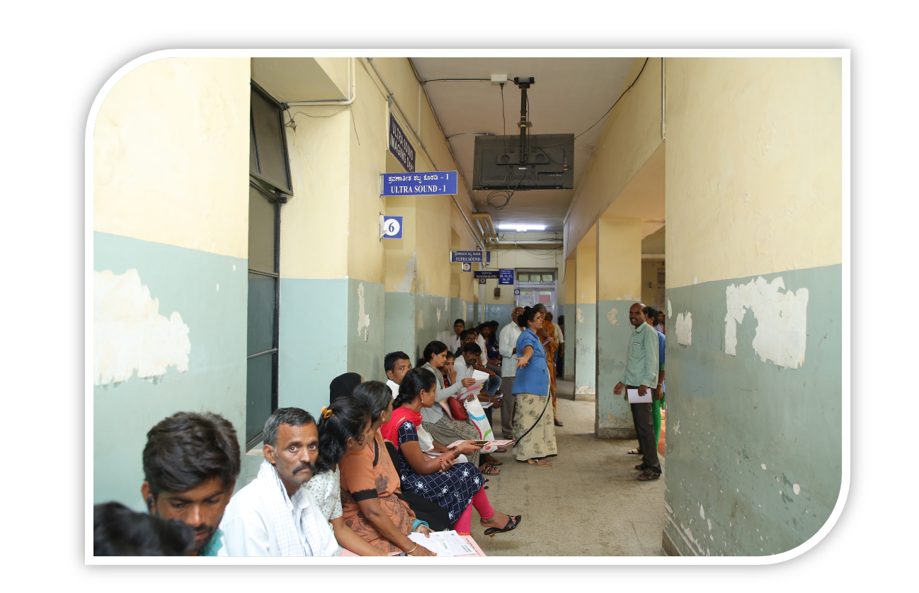
The Department has a full fledged Seminar room with a dedicated computer and LCD projector for teaching program for MD, B.Sc, M.Sc students. PACS room is present where cross section reporting is done. During the year patients are referred for Investigation from all over Karnataka State as well as from neighboring states. Tests are performed not only for malignant disorders, but also for non-malignant conditions. Institute patients, Staff Members and referral patients from other hospitals are being regularly investigated.
The department is staffed with well-qualified and trained personnel.
Academic Activities
- MCI recognized MD Radiodiagnosis course is being conducted in the department commencing from year 2019-20 two candidates per year are enrolled.
- RGUHS recognized fellowship programmes in Onco Imaging & Interventions is conducted for duration of 1 year with four candidates enrolled each year. This has started from 2019-20
- Senior Residents are allotted through Department of Health and Family welfare. Six posts are available for the same.
- RGUHS recongnized M.Sc course is conducted for duration of two years. 10 seats are allotted every year.
- Sc., medical Technology (Radiodiagnosis) Course-3 Years is run by the department. Several batches of Students are going through this course, which is of 3 Years duration.
Post Graduate, Under Graduate Students from various Medical Colleges are trained in various departments of Institute including Radiodiagnosis. Post Graduate students of various branches in the Institute Vis., M.ch, (Surgical oncology) MD., (Radiotherapy), Diploma in Nuclear medicine, DM., (Medical Oncology) Other B.Sc., Courses also attend regular postings in the department of Radiodiagnosis. Similarly MD and DMRD students from various MCI recognized institutes of Karnataka are posted on rotation basis in the department.
Radiologists and Technicians are given specialized training specifically in radiography, Ultrasonography, CT scanner, MRI & DSA.
Thrice weekly teaching program – Seminars/Case Presentation/journal clubs is undertaken in the department.
Interactive discussions within department and interdepartmental Residents and Faculty are mainstay of knowledge transfer.
Tumor Board Meetings are conducted with Department of Paediatrics ( Kidwai),Department of Paediatrics ( Indira Gandhi Child Care ), Head & neck Oncology , Medical Oncology on a regular basis. Department also participates in institute tumor boards and mortality meetings.
Work load in Department:
Department of Radiodiagnosis does minimum of nearly 200 radiographs, few special investigations such as barium studies, 60 ultrasound cases including 3-5 ultrasound neck and scrotum cases, 3 to 5 dopplers , 10-15 mammograms with sono mammography correlation, 60-70 CT scan per day. 20-30 MRI scans are done on a daily basis. Interventional Radiology cases including PTBD, TACE, Embolization Chemoport Insertion are being done about 30 to 50 in a month. Reviews of approximately 20-25 outside CT and MRI scans per day; upto 25 - 30 ultrasound guided FNAC, 4 to 6 biopsies per day and 4-6 CT guided FNAC / Biopsy every day are performed.
Major equipments added during the year
- PROGONOSIS –DR-Mobile Radiography -3 numbers
- ALLENGERS –CR-X-Rray -3 numbers
- PROGONOSIS –C-Arm
- SIEMENS – CT -64 SLICE – go-Up
Related Statistics of the Department
|
SL.NO
|
Types of Investigations
|
JAN
|
FEB
|
MAR
|
APRIL
|
MAY
|
JUN
|
JUL
|
AUG
|
SEP
|
OCT
|
NOV
|
DEC
|
Total
|
|
1.
|
X- rays & Radiography
|
1,852
|
1937
|
1774
|
1114
|
577
|
1291
|
2058
|
1,729
|
2019
|
1887
|
1784
|
1967
|
19,989
|
|
2.
|
Ultraosund
|
619
|
791
|
867
|
750
|
270
|
270
|
1273
|
1273
|
1289
|
805
|
936
|
1214
|
10,357
|
|
3.
|
U/SFNAC
|
301
|
298
|
252
|
117
|
107
|
107
|
261
|
287
|
275
|
311
|
250
|
272
|
2,838
|
|
4.
|
U/S BIOPSY
|
32
|
26
|
34
|
42
|
12
|
12
|
44
|
55
|
50
|
34
|
45
|
63
|
449
|
|
5.
|
Mammography
|
182
|
211
|
221
|
101
|
50
|
50
|
205
|
182
|
184
|
187
|
156
|
217
|
1,946
|
|
6.
|
U/S. Breast
|
18
|
24
|
16
|
11
|
14
|
14
|
18
|
8
|
11
|
15
|
25
|
27
|
201
|
|
7.
|
C.T.Scan
|
2521
|
3371
|
1038
|
733
|
797
|
797
|
1856
|
1944
|
3255
|
4099
|
4,556
|
2975
|
27,942
|
|
8.
|
CT FNAC
|
37
|
39
|
17
|
12
|
6
|
6
|
15
|
45
|
9
|
14
|
22
|
20
|
242
|
|
9.
|
CT Biopsy
|
72
|
45
|
62
|
56
|
20
|
20
|
56
|
78
|
40
|
60
|
32
|
96
|
637
|
|
10.
|
CT RT Planning
|
15
|
565
|
627
|
420
|
187
|
466
|
724
|
|
601
|
634
|
696
|
810
|
5,745
|
|
11.
|
MRI
|
572
|
290
|
353
|
258
|
140
|
374
|
636
|
608
|
450
|
670
|
620
|
850
|
5,821
|
|
12.
|
Echo
|
611
|
620
|
921
|
802
|
802
|
1003
|
1020
|
1180
|
1215
|
1320
|
1417
|
1376
|
12,287
|
|
13.
|
DSA
|
8
|
21
|
26
|
34
|
32
|
25
|
22
|
26
|
24
|
16
|
35
|
34
|
303
|
Conferences / Seminars/ Workshops conducted by the Department
Conference was conducted by the department on Contrast Media: TechAspire (12/11/2022)
Seminars conducted are as follows :
|
TOPIC
|
PRESENTER
|
|
Burkits lymphoma
|
Dr. Dilip
|
|
Wilms tumor
|
Dr. Dilip
|
|
GIST
|
Dr. Zain
|
|
Bone tumors
|
Dr. Zain
|
|
Ca.Prostate
|
Dr. Dilip
|
|
USG guided biopsy focal liver lesion
|
Dr. Zain
|
|
RCC
|
Dr. Dilip
|
|
TIRADS
|
Dr. Aiswarya Shankar Hugar
|
|
Congenital CNS injections
|
Dr. Sushma Sanagana Gouda Savadi
|
|
Metabolic bone disorder B-I
|
Dr. Sushma Sanagana Gouda Savadi
|
|
Metabolic bone disorder B-II
|
Dr. Kiran Budhihal
|
|
ILD-part -I
|
Dr. Aiswarya Shankar Hugar
|
|
ILD-part-II
|
Dr. Chetan.B
|
|
ILD-part-III
|
Dr. Chetan.B
|
|
GI Bleed and role of intervention
|
Dr. Sushma Sanagana Gouda Savadi
|
|
Radiological approach to the cyanotic heart disease
|
Dr. Kiran Budhihal
|
|
Aorta - congenital variations and anomalies
|
Dr. Chetan.B
|
|
CNS infections - Bacterial and tuberculosis
|
Dr. Aiswarya Shankar Hugar
|
|
Phakomatoses
|
Dr. Sushma Sanagana Gouda Savadi
|
|
Radiological features of arthropathies -I
|
Dr. Kiran Budhihal
|
|
Radiological features of arthropathies -II
|
Dr. Sushma Sanagana Gouda Savadi
|
|
Genitourinary Tract contrast imaging
|
Dr. Abhishek
|
|
Chest – X- ray film reading session
|
Dr. Prashanth
|
|
1ST trimester film reading session
|
Dr. Aiswarya Shankar Hugar
|
|
Abdominal X- ray reading session
|
Dr. Abhishek
|
|
Barium studies of GIT
|
Dr. Prashanth
|
|
Skull , Spine
|
Dr. Abhishek
|
|
II-I and weeks – obstetrics scan
|
Dr. Sushma Sanagana Gouda Savadi
|
|
GI embryology malrotation
|
Dr. Abhishek
|
|
TIFFA SCAN
|
Dr. Aiswarya Shankar Hugar
|
|
Genitourinary – Embroylosis and anomalies
|
Dr. Prashanth
|
|
Ultrasound assessment of fetal & growth
|
Dr. Sushma Sanagana Gouda Savadi
|
|
Female genital tract embryology and malformation
|
Dr. Abhishek
|
|
Obstetric Doppler and biophysical profile
|
Dr. Aiswarya Shankar Hugar
|
|
Skeletal dysplasia -I
|
Dr. Prashanth
|
|
Fetal ECHO
|
Dr. Sushma Sanagana Gouda Savadi
|
|
Congenital skeletal dysplasia II
|
Dr. Abhishek
|
|
Imaging post partum hemorrhage
|
Dr. Aiswarya Shankar Hugar
|
|
Ultrasound physics-I
|
Dr. Prashanth
|
|
Imaging in stroke
|
Dr. Aiswarya . K
|
|
RECIST & PET –RECIST
|
Dr. Divya
|
|
Sinonasal Ca. staging
|
Dr. Samanvita
|
|
ACR- BIRADS
|
Dr. Mayuresh
|
|
Staging & Reporting of Nasphryngeal Ca
|
Dr. Vivek
|
|
Carcinoma of tongue & base of tongue staging reporting
|
Dr. Venkateshwar
|
|
Staging and reporting Ca of the oral cavity
|
Dr. Zubair
|
|
Staging and reporting of tonsillar carcinoma
|
Dr. Revathi
|
|
Staging and reporting of soft hard palate carcinoma
|
Dr. Divya
|
|
BIRADS – MRI
|
Dr. Mayuresh
|
|
Staging and reporting of carcinoma hypopharm
|
Dr. Samanvita
|
|
Salivary gland tumor staging and reporting
|
Dr. Venkateshwar
|
|
Left hemithorax mass – IHC – Ewings sarcoma
|
Dr. Sachin
|
|
Thyroid Ca staging and reporting
|
Dr. Zubair
|
|
Staging & Imaging of neck glomus tumor of head & neck
|
Dr. Aiswarya .K
|
|
Staging and reporting of carotid body tumor
|
Dr. Samanvita
|
|
Lung RADS
|
Dr. Preetham
|
|
Endolymphatic sac tumor
|
Dr. Divya
|
|
Malignant PNST, Ewings sarcoma , mets from thyroid
|
Dr. Preetham
|
|
CT colonography exporting and data system (C- RADS)
|
Dr. Sachin
|
|
Reporting staging of Esophageal
|
Dr. Vivek
|
|
Neuroendocrine tumor , Carotid process tumor HPE correlation
|
Dr. Sachin
|
|
Staging and reporting of lung carcinoma
|
Dr. Venkateshwar
|
|
Left suprarenal gland benign nerve sheath tumor schwarnoma , Glomus jugulare
|
Dr. Aiswarya . K
|
|
Vesical imaging reporting and data system
|
Dr. Mayuresh
|
|
Reporting staging breast Ca
|
Dr. Zubair
|
|
Lung metastasis, Pontine glioma ,
Hemangioblastoma
|
Dr. Preetham
|
Research activities/projects/completed projects, ongoing and new projects
Thesis:
Comparitive Study Of American College Of Radiology Thyroid Imaging Reporting And Data System (ACR TIRADS) And Korean TIRADS With Pathological Correlation - Dr. Kiran.B.Budhihal
Comaprison Of Magnetic Resonnace Imaging And Trans Rectal Ultrasound In Clinically Suspected Prostatic Cancer Patients Undergoing Trans Rectal Ultrasound Guided Biopsy With Histopathological Correlation - Dr. Chetan .B
Role of Ultrasonography and Ultrasound elastography in the evaluation of thyroid nodules with cytopathological correlation – Dr. Aiswarya Shankar Hugar
Quantitative elastography combined with BIRAD’S in differentiating benign and malignant solid breast lesion with histopathology as gold standard - Dr. Sushma Sanganna Gouda Savadi
Role of Neck Imaging reporting and data system in the detection of local regional recurrence of head and neck squamous cell cancers by cross sectional imaging modalities - Dr. Abhishek
Role of radiofrequency ablation in hepatic lesions and comparing the efficacy of radiofrequency ablation in lesions less than 3 cms and lesions 3-5 cms in achieving complete response - Dr. Prashanth
Staff Details
Dr. Madhu.SD: Professor & HOD
Dr. Anuradha Kapali: Associate Professor
Dr. Suhas.B: Assistant Professor
Dr. Shrunga Tejaswi : Assistant Professor ( ADHOC)
Dr. Dhanwin.R.Shetty : Assistant Professor ( ADHOC)
Dr. Revathi.V : Senior Resident ( ADHOC)
Dr.Syed Zubair Ahmed : Senior Resident ( ADHOC)
Dr.Divya G.A:Senior Resident ( ADHOC)
Dr.Venkateswar.K.V:Senior Resident ( ADHOC)
Dr.Vivek :Senior Resident ( ADHOC)
Dr. Samanvita Gode :Senior Resident ( ADHOC)
Mr. Sudhir.N, Senior Medical Imaging Technologist
Mr. Siddalingeswar.H.V, Senior Medical Imaging Technologist
Mr. Javaraj.N, Senior Medical Imaging Technologist
Mr.Satya Prasad, Senior Medical Imaging Technologist
Mr.D. Anantha Raman, Senior Medical Imaging Technologist
Mr. Maheswara Murthy .M, Senior Medical Imaging Technologist
Mr. Sidhara K.A, Chief Medical Imaging Technologist
Mr. Darshan. B.S, Medical Imaging Technologist
Mr. Chandrakanth.K.S, Medical Imaging Technologist
Mr. Praveen. S.U, Medical Imaging Technologist
Mr. Vinay Gowda, Medical Imaging Technologist
Mr. Vikas, Medical Imaging Technologist
Ms. Yashaswini .R. Gowda, Medical Imaging Technologist
Mr. Srinivasa.R, Medical Imaging Technologist
Ms.Ganavi.R, Medical Imaging Technologist
Ms.Jyothi .S, ECHO Technologist
Mrs. Kamakshi.B, Data Entry Operator
Mrs. Harshitha, Data Entry Operator
Mr. Ravi Kumar .M, Data Entry Operator
Mrs. Nalini Siddappa, Data Entry Operator
Mr. Hitesh.K., Data Entry Operator
Mr. Umesh, Grop’’D”Attender
Papers published in National and International Journals.
Madhu SD, Parvathi M, Jyothsna Rani, Sujatha Patnaik. MELIOIDOSIS: A RARE BUT EMERGING INFECTIOUS DISEASE IN INDIA AND ROLE OF RADIOLOGIST IN DIAGNOSIS. International Journal of Anatomy, Radiology and Surgery .
Madhu SD, Jaipal R Beerappa, Pooja, RaghuRam P Role of DWI in Detection and Characterization of Focal Liver Lesions International Journal of Anatomy, Radiology and Surgery.
Madhu SD, Jaipal, Prashanth Sinha, Raghuram Role of MR Spectroscopy in Differentiating Tumor Recurrence and Post Radiation Changes in Treated Brain Tumors with Radiotherapy, International Journal of Anatomy, Radiology and Surgery.
Papers published in National and International Journals (Archive)
Madhu SD, Parvathi M, Jyothsna Rani, Sujatha Patnaik. MELIOIDOSIS: A rare but emerging infectious disease in india and role of radiologist in diagnosis. International Journal of Anatomy, Radiology and Surgery.
Madhu SD, Jaipal R Beerappa, Pooja, RaghuRam P Role of DWI in Detection and Characterization of Focal Liver Lesions International Journal of Anatomy, Radiology and Surgery
Madhu SD, Jaipal, Prashanth Sinha, Raghuram Role of MR Spectroscopy in Differentiating Tumor Recurrence and Post Radiation Changes in Treated Brain Tumors with Radiotherapy, International Journal of Anatomy, Radiology and Surgery.
Madhu SD, Parvathi M, Divya Lakshmi, Sneha Volvoikar Isolated involvement of cervical lymph nodes in Castleman’s disease in a young patient: A rare presentation J. Evolution Med. Dent. Sci./eISSN- 2278-4802, pISSN- 2278-4748/ Vol. 05/ Issue 39/ May 16, 2016.
Role of Ultrasonography in Thyroid Nodules with Pathological correlation. International Journal of contemporary Medical research.2016/ Vol 3/ Issue 5/ Page 1451-53.
Anuradha Kapali, Jaipal B R, Raghuram.P, Ravindra Bangar, Sateesh Kumar Atmakuri. Role of ultrasonography in thyroid nodules with pathological correlation. International Journal of Contemporary Medical Research 2016;3(5):1451-1453.
Role of MRI in staging of carcinoma Cervix. Journal of evidence based medicine and healthcare. 2016/ Vol 3/ Issue 31/ Page 1396-1400.
Beerappa JR, Kapali A, Pream C, et al. Role of MRI in staging of carcinoma cervix. J. Evid. Based Med. Healthc. 2016; 3(31), 1396-1400. DOI: 10.18410/jebmh/2016/320.
Diagnostic accuracy of Ultrasound imaging in Hashimoto’s thyroiditis. Thyroid Research and practice. 2017/ Vol 14/ issue 1/ Page 28-31
Kapali A, Beerappa J, Raghuram P, Bangar R. Diagnostic accuracy of ultrasound imaging in Hashimoto’s thyroiditis. Thyroid Res Pract 2017;14:28-
Mammographic and sonomammographic evaluation of breast masses with Pathological correlation : A prospective original study. International Journal of Anatomy, Radiology and Surgery. 2016 Jul / Vol-5(3) / RO09-RO12
Jaipal R Beerappa, Balu S, Nandan Kumar L D, Anuradha Kapali, Raghuram P International Journal of Anatomy, Radiology and Surgery. 2016 Jul, Vol-5(3): RO09-RO12.
Unilateral renal cystic disease : A case report Journal name : Journal of evolution of medical and dental sciences. Case report. 2015/ Vol 4/ Issue 56/ Page 9839-9842.
Anuradha Kapali, P. Raghuram, Beerappa Jaipal, Sateesh Kumar Atmakuri, Ravindra Bangar. “Unilateral Renal Cystic Disease: A Case Report”. Journal of Evolution of Medical and Dental Sciences 2015; Vol. 4, Issue 56, July 13; Page: 9838-9841, DOI:10.14260/jemds/2015/1420.
carcinoma cervix with fat attenuating skull metastases. Journal of cancer metastases and treatment. 2016 / Vol 2 / Page 228-30.
Kapali A, Kumar AS, Malathi M, Shamsundar SD. Carcinoma cervix with fat attenuating skull metastases. J Cancer Metastasis Treat 2016;2:228-30.
Hepatic angiomyolipoma: A radiological dilemma. Oncology, gastroenterology and hepatology reports. 2016 / Vol 5 / Issue 2 / Page 60-62.
Kapali A, Jaipal BR, Parampalli R, Atmakuri SKumar. Hepatic Angiomyolipoma: A Radiological Dilemma. Oncology, Gastroenterology and Hepatology Reports. 2016;5(2):60-62.
Dr. ATHIRA. Diffusion weighted imaging in breast cancer - Can it be a noninvasive predictor of nuclear grade? Indian Journal of Radiology and Imaging.2020 Jan-Mar; 30(1): 13–19.
Pre operative contrast enhanced computer tomographic evaluation of cervical nodal metastatic disease in oral squamous cell carcinoma – Page 310, Indian Journal of Cancer (Oct-Dec 2013) 50Role of MR spectroscopy in differentiating tumor recurrence and post radiation changes in treated brain tumors with radiotherapy.International Journal of Anatomy, Radiology and Surgery. 2016 Jul, Vol-5(3): RO52-RO58.
Madhu SD,Dr. Jaipal R. Beerappa, Dr. Prashanth KumarSinha,Dr. Raguram.P.Role of DWI in detection and characterization of focal liver lesions. International Journal of Anatomy, Radiology and Surgery. 2016 Jul, Vol-5(3): RO59-RO66.
Madhu SD, Jaipal R Beerappa, Pooja, RaghuRam.P Role of ultrasonography in thyroid nodules with pathological correlation. International Journal of Contemporary Medical Research 2016;3(5):1451-1453.
Anuradha Kapali, Jaipal B R, Raghuram.P, RavindraBangar, Sateesh Kumar Atmakuri. Role of MRI in staging of carcinoma cervix. J. Evid. Based Med. Healthc. 2016; 3(31), 1396-1400. DOI: 10.18410/jebmh/2016/320.
Beerappa JR, Kapali A, Pream C, et al.Diagnostic accuracy of Ultrasound imaging in Hashimoto’s thyroiditis. Accepted for publication. Thyroid Research and practice. Mammographic and sonomammographic evaluation of breast masses with Pathological correlation: A prospective original study. International Journal of Anatomy, Radiology and Surgery. 2016 Jul, Vol-5(3): RO09-RO12
Jaipal R Beerappa, Balu S, Nandan Kumar L D, Anuradha Kapali, Raghuram P “Unilateral Renal Cystic Disease: A Case Report”. Journal of Evolution of Medical and Dental Sciences 2015; Vol. 4, Issue 56, July 13; Page: 9838-9841, DOI:10.14260/jemds/2015/1420.
Anuradha Kapali, P. Raghuram, Beerappa Jaipal, Sateesh Kumar Atmakuri, Ravindra Bangar. Carcinoma cervix with fat attenuating skull metastases. J Cancer Metastasis Treat 2016;2:228-30.
Kapali A, Kumar AS, Malathi M, Shamsundar SD. Hepatic Angiomyolipoma: A Radiological Dilemma. Oncology, Gastroenterology and Hepatology Reports. 2016;5(2):60-62.
Kapali A, Jaipal BR, Parampalli R, AtmakuriSKumar. Anuradha Rao , Divya Lakshmi, Raghuram.P (2017, Sep. 28) Spontaneous Reno-colic Fistula As Initial Presentation Of Renal Cell Carcinoma {Online}URL: http://www.eurorad.org/case.php?id=15065.
Rao A, Sharma C, Parampalli R. Role of diffusion-weighted MRI in differentiating benign from malignant bone tumors 2019; 1: 20180048.Original article in BJR/Open ; https:// doi. org/ 10. 1259/ bjro. 20180048.
Madhu SD, Parvathi M, Jyothsna Rani, Sujatha Patnaik. MELIOIDOSIS : A RARE BUT EMERGING INFECTIOUS DISEASE IN INDIA AND ROLE OF RADIOLOGIST IN DIAGNOSIS. International Journal of Anatomy, Radiology and Surgery
Madhu SD, Jaipal R Beerappa, Pooja, RaghuRam P Role of DWI in Detection and Characterization of Focal Liver Lesions International Journal of Anatomy, Radiology and Surgery
Madhu SD, Jaipal, Prashanth Sinha, Raghuram Role of MR Spectroscopy in Differentiating Tumor Recurrence and Post Radiation Changes in Treated Brain Tumors with Radiotherapy, International Journal of Anatomy, Radiology and Surgery.
Madhu SD, Parvathi M, Divya Lakshmi, Sneha Volvoikar Isolated involvement of cervical lymph nodes in Castleman’s disease in a young patient: A rare presentation J. Evolution Med. Dent. Sci./eISSN- 2278-4802, pISSN- 2278-4748/ Vol. 05/ Issue 39/ May 16, 2016.
Anuradha Kapali, Jaipal B R, Raghuram P, Ravindra Bangar, Sateesh Kumar Atmakuri. Role of ultrasonography in thyroid nodules with pathological correlation. International Journal of Contemporary Medical Research 2016;3(5):1451-1453.
Anuradha Kapali, Beerappa J, Raghuram P, Bangar R. Diagnostic accuracy of ultrasound imaging in Hashimoto’s thyroiditis. Thyroid Res Pract 2017;14:28-31.
Anuradha Kapali, P. Raghuram, Beerappa Jaipal, Sateesh Kumar Atmakuri, Ravindra Bangar. “Unilateral Renal Cystic Disease: A Case Report”. Journal of Evolution of Medical and Dental Sciences 2015; Vol. 4, Issue 56, July 13; Page: 9838-9841, DOI:10.14260/jemds/2015/1420.
Anuradha Kapali A, Kumar AS, Malathi M, Shamsundar SD. Carcinoma cervix with fat attenuating skull metastases. J Cancer Metastasis Treat 2016;2:228-30.
Anuradha Kapali A, Jaipal BR, Parampalli R, Atmakuri SKumar. Hepatic Angiomyolipoma: A Radiological Dilemma. Oncology, Gastroenterology and Hepatology Reports. 2016;5(2):60-62.
Patil DD, Pujalwar S. Singh YD. Hallervorden Spatz disease. J. Evolution Med. Dent. Sci. 2018;7(52): 5570-5572, DOI: 10.14260/jemds/2018/1232.
Rao A, Sharma C, Parampalli R. Role of diffusion-weighted MRI in differentiating benign from malignant bone tumors 2019; 1: 20180048.Original article in BJR/Open ; https:// doi. org/ 10. 1259/ bjro. 20180048.